Radiologic Approach to Erectile Dysfunction. Main causes, diagnosis and therapeutic options in interventional radiology

Buy Lab Tests Online

madman

Super Moderator
Learning objectives
  • Learn about erectile dysfunction. Pathophysiology, symptoms and main causes.
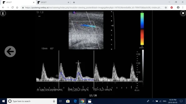
  • Stimulated Color Doppler ultrasound diagnostic criteria. Arteriogenic erectile dysfunction, veno-occlusive erectile dysfunction , mixed aetiology and false venous-leak.
  • Role of interventional radiology in the treatment of venous-leak.
  • Endovascular embolisation therapy.


Background

Erectile dysfunction (ED) is defined as the inability to attain or maintain a penile erection of sufficient quality to allow satisfactory sexual performance.

The prevalence of this condition increases with age with a 39% prevalence at the age of 40 years and a 67% prevalence at the age of 70 years. It has been estimated that the worldwide prevalence of erectile dysfunction will be 322 million cases by the year 2025.


Fig. 1: Physiology of Penile Erection.
NO: Nitric Oxide. GTP:guanosine triphosphate PDE5 phosphodiesterase type 5. cGMP: cyclic guanosine monophosphate.
References: Radiology, Hospital Fundación Jiménez Díaz, Hospital Universitario Fundación Jiménez díaz - Madrid/ES
Screenshot (77).png


Fig. 2: Veno-occlusive mechanism in penile erection. A- Normal flacid penis. Smooth muscle cells are contracted and the sinusoid spaces in corpus cavernosum are empty. B- Normal erect penile anatomy. The increase of NO levels allows smooth muscle cells relaxation, sinusoid spaces are enlarged and compress the subtunical venous plexus. C-Several mechanism of venogenic erectile dysfunction.
References: McVary KT. Erectile Dysfunction. N Engl J Med 2007;357:2472-81.
Screenshot (78).png


Fig. 3: Main Causes of Erectile Dysfunction.
Screenshot (79).png


Fig. 4: Risk Factors in Erectile Dysfunction. Endothelial Dysfunction as a shared physiopathological mechanism between ED and cardiovascular diseases.
References: Radiology, Hospital Fundación Jiménez Díaz, Hospital Universitario Fundación Jiménez díaz - Madrid/ES
Screenshot (80).png


Fig. 5: The Hypothesis of Arterial Diameter. - Given coronary arteries diameter, myocardial vascularisation would be affected secondarily after penile vascularisation. - Coronary Arteries Diseases and Erectile Dysfunction:different clinical signs of same progressive disease.
References: Montorsi P et al. (2003). Eur Urol ; 44: 352-354.
Screenshot (81).png
 
Defy Medical TRT clinic doctor

madman

Super Moderator
Findings and procedure details


Screenshot (82).png

Fig. 6: Normal anatomy of penis. CC: Corpora cavernosa. CE: Corpus spongiosum. Arrowheads: Tunica Albuginea. Stars: Cavernosal arteries. Arrow: Buck's fascia.
References: Radiology, Hospital Fundación Jiménez Díaz, Hospital Universitario Fundación Jiménez díaz - Madrid/ES


Screenshot (83).png

Fig. 7: Schematic view of arterial vascularization of the penis. The dorsal artery of penis, cavernosal arteries and urethral artery are brancehs of the internal pudendal artery (IPA).
References: Philip F, Shishebor MH. Current State of Endovascular Treatment for Vasculogenic Erectile Dysfunction.Curr Cardiol Rep (2013) 15:360.


Screenshot (84).png

Fig. 8: Vascular Anatomy of penis.
References: Baxter GM and Sidhu PS. Ultrasound of the Urogenital System. Thieme, Stuttgart, 2006, Ch. 12.


Screenshot (85).png

Fig. 9: PGE-1 Stimulated CDUS of penis in Erectile Dysfunction Tecnique. Left: Injection of 20 micrograms of PGE-1 with 30 needle inside the corpora cavernosa. Right: Colour flow Doppler ultrasound of the cavernosal arteries should be performed with the probe positioned at the base of the penis on the ventral surface. The angle for Doppler analysis needs to be optimised (<60°) with box-steering, angle correction and orientation of the probe to ensure reproducible, valid measurements.
References: Berrkhim BM. Doppler Duplex Ultrasonography of the Penis. J Sex Med 2016;13:726e731


Screenshot (86).png

Fig. 10: CDUS of right cavernosal artery before injection of PGE-1. Artery diameter less than 1 mm and PSV <10 cm/s.
References: Radiology, Hospital Fundación Jiménez Díaz, Hospital Universitario Fundación Jiménez díaz - Madrid/ES
 

madman

Super Moderator
Screenshot (87).png

Fig. 11: CDUS of left cavernosal artery after PGE-1 injection. PSV of 30 cm/s, EDV is absent or even reversal. Both hemodynamic parameters indicates normal response after PGE-1 stimulation.


Screenshot (88).png

Fig. 12: Stimulated CDUS after PGE-1 injection. Arterial Insufficiency. PSV of 20,3 cm/s, EDV of 3,4 cm/s, RI<0,75.
References: Radiology, Hospital Fundación Jiménez Díaz, Hospital Universitario Fundación Jiménez díaz - Madrid/ES


Screenshot (89).png

Fig. 13: Stimulated CDUS of left cavernosal artery. Veno-occlusive (venous-leak) dysfunction. PSV of 30 cm/s, EDV of 10,3 cm/s, RI 0,82.
References: Radiology, Hospital Fundación Jiménez Díaz, Hospital Universitario Fundación Jiménez díaz - Madrid/ES


Screenshot (90).png

Fig. 14: Stimulated CDUS after PGE-1 injection. Mixed aetiology of erectile dysfunction. PSV 22,8 cm/s, EDV 7,7 cm/s, RI 0,66.
References: Radiology, Hospital Fundación Jiménez Díaz, Hospital Universitario Fundación Jiménez díaz - Madrid/ES


Screenshot (91).png

Fig. 15: Haemodynamic Parameters for Stimulated CDUS in ED.
References: Radiology, Hospital Fundación Jiménez Díaz, Hospital Universitario Fundación Jiménez díaz - Madrid/ES
 

Attachments

  • Screenshot (88).png
    Screenshot (88).png
    589.5 KB · Views: 141

madman

Super Moderator
Screenshot (92).png

Fig. 16: Caverno-CT images of 4 patients with ED. Anomalous Venous Drainage Classification. A- Type A (no leakage). The cavernous bodies are both well opacified, without opacification of superficial vein either deep dorsal vein or opacification of the periprostatic plexus. B- Type B leakage. The cavernous bodies are both opacified, with the deep dorsal vein and internal pudendal veins alone indicating a type B leakage. C- Type C leakage. The cavernous bodies are partially opacified, with an obvious opacification of the superficial dorsal vein draining in the right great saphena vein, responsible for a type C leakage. D- Type D leakage: The cavernous bodies are opacified, with an opacification of the two superficial dorsal veins draining in the right femoral vein, and also an opacification of the two deep dorsal veins and internal also pudendal veins responsible for a type D leakage.
References: Virad R, Paul JF.New Classification of Anomalous Venous Drainage Using Caverno-Computed Tomography in Men with Erectile Dysfunction.J Sex Med 2011; 8:1439-1444.


Screenshot (93).png

Fig. 17: Cavernography-CT based Classification of Anomalous Venous Drainage in Erectile Dysfunction.
References: Radiology, Hospital Fundación Jiménez Díaz, Hospital Universitario Fundación Jiménez díaz - Madrid/ES

Screenshot (94).png

Fig. 18: Schematic view of the complex venous drainage of the penis.
References: Rebonato A et al. Embolization of the Periprostatic Venous Plexus for Erectile Dysfunction Resulting from Venous Leakage. J Vasc Interv Radiol 2014; 25:866–872.

Screenshot (95).png

Fig. 19: Case 1. Patient with veno-oclusice erectile dysfunction. A- Cavernography after PGE-1 injection in corpora cavernosa reveals a type B venous-leakage at he level of deep pelvic veins/periprostatic venous plexus on both sides (arrows in B) B- Type B venous leakage with abnormal outflow towards deep pelvic veins and periprostatic venous plexus bilaterally (Arrows). C- superselective retrograde venous angiography of right sided dilated venous periprostatic plexus and embolisation with coil (shor arrow) and glue (histoacryl-lipiodol)(long arrow). D-Control cavernography after complete embolisation of both sides with filling of the target vessel system. There is no venous-leakage. Technical success was achieved.
References: Radiology, Hospital Fundación Jiménez Díaz, Hospital Universitario Fundación Jiménez díaz - Madrid/ES


Screenshot (96).png

Fig. 20: Case 2. Patient with veno-oclusive erectile Dysfunction with CDUS suggestive of venous-leak. A- Cavernography after PGE-1 injection in corpora cavernosa shows a type C venous-leakage towards right external pudendal and great saphenous vein (arrow). B - Superselective retrograde venous angiography of right external pudendal vein (arrow). C- Embolisation with 1 coil and glue (histoacryl-lipiodol) up to complete filling of target vessel (arrow). Notice the radiopaque appearance of the Lipiodol to control the result of embolisation. D- Venous access in deep dorsal vein with micropuncture set. E- 4 months later Control anterograde venous angiography using the deep dosal vein access. Notice the complete occlusion of the right external pudendal vein and recidive of venous-leak towards deep prostatix plexus. Technical success was achieved.
References: Radiology, Hospital Fundación Jiménez Díaz, Hospital Universitario Fundación Jiménez díaz - Madrid/ES
 

madman

Super Moderator
Conclusion
Erectile dysfunction is defined as the inability to attain or maintain a penile erection of sufficient quality to allow satisfactory sexual performance. The prevalence of this condition increases with age.

There are many causes of erectile dysfunction including organic, psychogenic, and combined causes. It is essential to identificate organic causes, because they are potentially treatables.

Erectile dysfunction is now being recognized as one of the earliest manifestations of endothelial dysfunction and peripheral vascular disease.

Stimulated CDUS of cavernosal arteries is the initial imaging technique of election to assess ED from vascular origin. A PSV> 30 cm/s and an RI higher than 0.8 are generally considered normal. A EDV >5cm/s is suggestive of venous-leak.

Transcatheter venous embolisation for veno-occlusive dysfunction in ED is a safe and effective therapeutic option. The minimally invasive nature of the procedure enabled us to perform the procedure in an out-patient setting with low costs. The ability to occlude even tiny venous collaterals that can be overlooked during surgical ligation makes this procedure superior to surgical treatment.
 

Attachments

  • APR5-ECR2019_C-3026.pdf
    4.4 MB · Views: 150
Last edited:
Buy Lab Tests Online
Defy Medical TRT clinic

Sponsors

cheap enclomiphene
nelson vergel coaching for men
Discounted Labs
TRT in UK Balance my hormones
Testosterone books nelson vergel
Register on ExcelMale.com
Trimix HCG Offer Excelmale
Thumos USA men's mentoring and coaching
Testosterone TRT HRT Doctor Near Me

Online statistics

Members online
3
Guests online
6
Total visitors
9

Latest posts

bodybuilder test discounted labs
Top